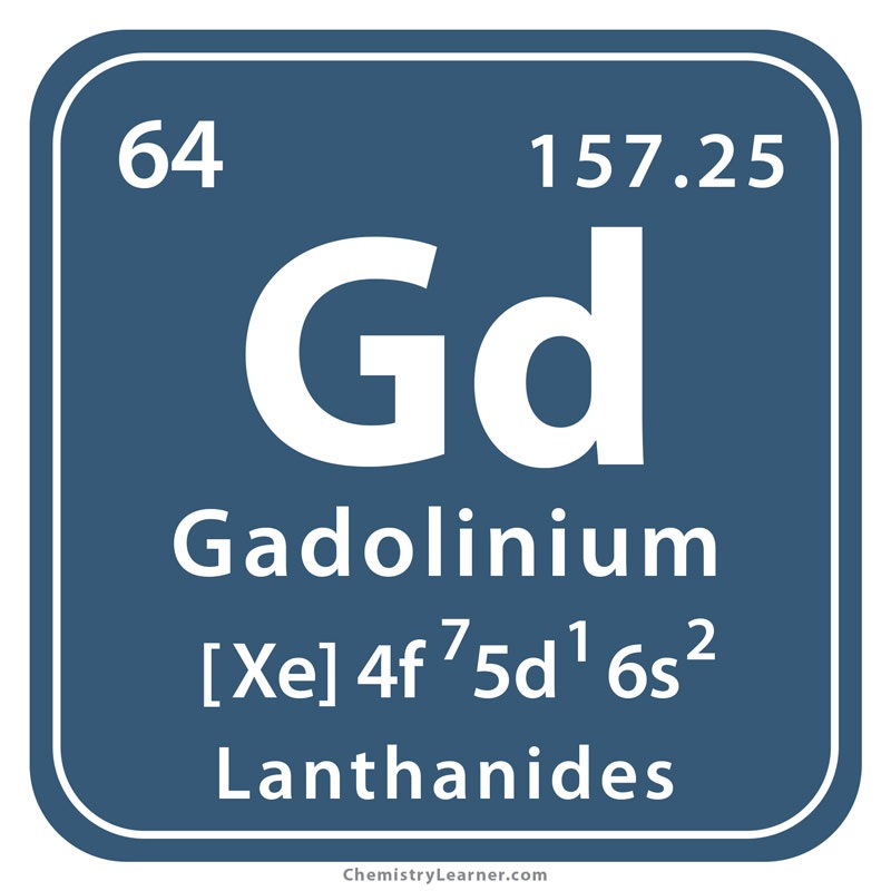
In a new study published in Scientific Reports , a team led by Brent Wagner, MD, MS, associate professor in the UNM Department of Internal Medicine, describes the use of electron microscopy to detect small deposits of gadolinium in the kidneys of women. people who had been injected with contrast agents before their MRIs.
“These are nanoparticles,” Wagner said. "They’re actually forming nanomaterial inside these cells." Gadolinium-based contrast agents were first introduced in the 1990s when MRI studies became more routine. Gadolinium aligns with the powerful magnetic field of an MRI scanner, resulting in sharper images, but because of its toxicity, the metal must be tightly bound to chelating molecules so it can be filtered through the kidneys and eliminated. .
But researchers have found that some gadolinium atoms can leak from contrast agents into the kidneys and other tissues, Wagner said. The effect was found in both rodents and humans, she said.
Gadolinium-containing contrast agents are used in about 50% of MRIs, Wagner said. An important question is why some people develop the disease, but most exposed people never develop negative symptoms.
An additional concern is that gadolinium appears to be reaching the environment. Because the MRI contrast agent is passed through urine, it is released into sewage systems, but wastewater treatment plants are not equipped to remove it.
The properties of contrast agents for magnetic resonance imaging (MRI) are based on a rare earth metal, gadolinium . Because gadolinium is toxic, MRI contrast agents are proprietary aminopolycarboxylic acid chelates designed to bind the metal tightly and improve renal clearance. Complications of MRI contrast agents include gadolinium encephalopathy, acute kidney injury, gadolinium deposition disease/symptoms associated with gadolinium exposure (SAGE), and ’nephrogenic’ systemic fibrosis. Exposure to any class of MRI contrast agent leads to long-term retention of gadolinium. Residual gadolinium from MRI contrast agent exposure has been found in all vital organs, including the brain, in both patients and animal models. Urine may contain gadolinium years after exposure to MRI contrast agents.
Our rodent models demonstrated the formation of gadolinium-rich nanoparticles in the kidney and skin after systemic treatment with MRI contrast agent. Gadolinium-rich densities have been found in the neuronal cytoplasm and nuclei of the brains of people exposed to MRI contrast agents during the course of routine care. The nanotoxicological mechanisms of gadolinium-induced disease are poorly understood. Our understanding of MRI contrast-induced complications is far from complete. These studies were performed to characterize the composition of intracellular gadolinium-rich minerals that form after systemic treatment with MRI contrast agents.
The onset of rare earth metallosis begins with renal gadolinium-rich nanoparticles from exposure to magnetic resonance imaging contrast agents. Summary The leitmotifs of magnetic resonance imaging (MRI) contrast agent-induced complications range from acute kidney injury, symptoms associated with gadolinium exposure (SAGE)/gadolinium deposition disease, life-threatening gadolinium encephalopathy, and irreversible systemic fibrosis . Gadolinium is the active ingredient in these contrast agents, a non-physiological lanthanide metal . The mechanisms of MRI contrast agent-induced diseases are unknown. Mice were treated with an MRI contrast agent. Human kidney tissues were obtained and analyzed from contrast-naïve and MRI contrast agent-treated patients. Kidneys (human and mouse) were evaluated with transmission electron microscopy and scanning transmission electron microscopy with X-ray energy dispersive spectroscopy. Treatment with magnetic resonance contrast agent resulted in unilamellar vesicles and mitochondriopathy in the epithelium. renal. Electron-dense intracellular precipitates and the outer edge of lipid droplets were rich in gadolinium and phosphorus. We conclude that MRI contrast agents are not physiologically inert . The long-term safety of these synthetic metal-ligand complexes, especially with repeated use, needs to be further studied. |
Discussion and Conclusions
Our results suggest that gadolinium is released from MRI contrast agent formulations in vivo and metabolized into mineralized intracellular nanoparticles. The high concentrations of phosphorus (and oxygen) suggest that the nanoparticles contain insoluble GdPO4 (and perhaps Gd2O3/Gd (OH)3) or a more complex/heterogeneous mineral. The phosphorus deposit is unknown. The abundance of phosphorus in lipids and the systemic response to gadolinium suggest that leaching from intracellular membranes may be one mechanism.
Gadolinium is not a physiological element. It is reasonable to assume that iatrogenic kidney injury, systemic fibrosis, dermal plaques, and SAGE are part of a spectrum of disorders that result from the retention of a toxic lanthanide metal. Nanotoxicity is undoubtedly a mediator of contrast agent complications in MRI. Differential breakdown of MRI contrast agents may explain susceptibility to complications.


