Summary Patients undergo interventions to achieve a "normal" brain temperature; a parameter that remains undefined for humans. The profound sensitivity of neuronal function to temperature implies that the brain should be isothermal, but observations of patients and non-human primates suggest significant spatiotemporal variation. Our objective was to determine the clinical relevance of brain temperature in patients by establishing how much it varies in healthy adults. We retrospectively examined data from all patients recruited to the High Resolution Intensive Care Unit Substudy of the European Collaborative Investigation into the Effectiveness of Neurotrauma in Traumatic Brain Injury (CENTER-TBI). Only patients with direct brain temperature measurements and without specific temperature control were included. To interpret patient analyses, we prospectively recruited 40 healthy adults (20 men, 20 women, 20–40 years) for brain thermometry using magnetic resonance spectroscopy. Participants were scanned in the morning, afternoon, and evening of a single day. In patients (n = 114), brain temperature ranged from 32.6 to 42.3 °C and mean brain temperature (38.5 ± 0.8 °C) exceeded body temperature (37.5 ± 0.8 °C). 5 °C, P < 0.0001). Of 100 patients eligible for brain temperature rhythm analysis, 25 showed a daily rhythm and the range of brain temperature decreased in older patients (P = 0.018). In healthy participants , brain temperature ranged from 36.1 to 40.9°C; Mean brain temperature (38.5 ± 0.4 °C) exceeded oral temperature (36.0 ± 0.5 °C) and was 0.36 °C higher in luteal females relative to females and males. follicular males (P = 0.0006 and P < 0.0001, respectively). Temperature increased with age , especially in deep brain regions (0.6°C over 20 years, P = 0.0002) and varied spatially by 2.41 ± 0.46°C with the highest temperatures in the thalamus. Brain temperature varied by time of day , especially in deep regions (0.86°C, P = 0.0001), and was lowest at night. From the healthy data we created HEATWAVE, a 4D map of human brain temperature. Testing the clinical relevance of HEATWAVE in patients, we found that the lack of a daily brain temperature rhythm increased the odds of death in intensive care 21-fold (P = 0.016), while absolute maximum or minimum temperature did not predict the outcome. However, warmer mean brain temperature was associated with survival (P = 0.035), and aging by 10 years increased the odds of death 11-fold (P = 0.0002). Human brain temperature is higher and varies more than previously assumed, depending on age, sex, menstrual cycle, brain region, and time of day. This has important implications for temperature control and management, as daily brain temperature rhythmicity emerges as one of the strongest individual predictors of survival after brain injury. We conclude that rhythmic daily variation in brain temperature, not absolute brain temperature, is one way in which human brain physiology can be distinguished from pathophysiology. |
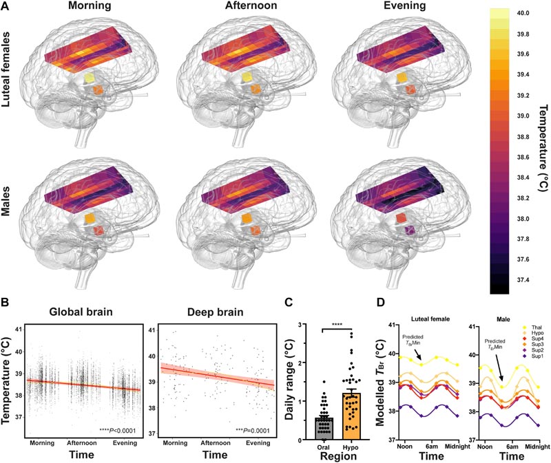
Figure: Healthy brain temperature varies depending on the time of day. (A) Snapshot of 3D TBr maps at each data collection point. The Inferno color scale is used to assign a temperature to each tissue voxel, with a resolution of 0.1°C. Aggregated temperatures are shown at each voxel for luteal females (n = 14) and males (n = 20) separately. (B) Linear mixed model results for TBr by time of day; Results for global TBr (left) and deep brain TBr (thalamus and hypothalamus, right) are shown. Solid red lines represent model fits, shaded areas represent 95% CIs, dark gray circles show residuals (single temperature data points), and smoothed dashed yellow lines represent partial residuals. The x-axis for time summarizes the continuous variable of time distance from the participant’s MSFsc (proportion of a linearized unit circle, where 0 = MSFsc and 1 = 24 h). (C) Temperature range (maximum vs. minimum at three time points tested) for oral and hypothalamic sites for each healthy participant (n = 39). Temperature varied more by time of day in the hypothalamus than orally (one-way repeated measures ANOVA with Sidak’s multiple comparisons test ****P < 0.0001; see Supplementary Fig. 10B for other brain regions). (D) Scheme for modeling 24-h temperature rhythms of the healthy human brain. Extrapolated TBr rhythms in healthy luteal women (n = 14) and men (n = 20), without controlling for age, BMI, or chronotype. Extrapolated temperature rhythms were created by doubling the mean temperatures measured at three time points and applying a 24-h sinusoidal fit to these six points. Note the higher temperatures in all regions in luteal females relative to males and the marked variation in deep brain temperatures in males. Arrows point to predicted TBr minima around 2-3 am (approaching MSFsc). Sup1–4 = superficial brain regions 1–4 from medial to lateral; hypo = hypothalamus; Thal = thalamus.
Discussion
We have established a 4D map of human TBr and show how this parameter varies with time of day, brain region, age and sex in adults. Human brain tissue clearly functions normally at temperatures 1 to 3 °C higher than generally assumed ; This discovery alone has challenging implications for neurocritical care. These data provide clinicians with an urgently needed, easily accessible reference resource for evidence-based interpretation of TBr data in patients.
Finally, we have found that a daily TBr rhythm is associated with a 21-fold higher probability of survival after brain injury, illustrating the high prognostic value of time-resolved TBr measurements and powering a prediction of mortality based on temperature.
Overall these results indicate that daily TBr rhythms are strongly associated with healthy brain function and are progressively compromised with age. Future studies should address whether supporting normal TBr variation is beneficial to patients.
Comments
Study links a daily cycle in brain temperature to survival after traumatic brain injury.
New research has shown that normal human brain temperature varies much more than we thought, and this could be a sign of healthy brain function. In healthy men and women, in whom the oral temperature is usually below 37 °C, the average brain temperature is 38.5 °C, and the deeper brain regions usually exceed 40 °C, especially in women during the day.
Previously, studies of human brain temperature relied on capturing data from brain-injured patients in intensive care, where direct brain monitoring is often needed. More recently, a brain scanning technique, called magnetic resonance spectroscopy (MRS), has allowed researchers to measure brain temperature non-invasively in healthy people. However, until now, MRS has not been used to explore how brain temperature varies throughout the day, or to consider how it influences an individual’s "body clock . "
The new study, led by researchers at the Medical Research Council (MRC) Laboratory of Molecular Biology, in Cambridge, UK, has produced the first 4D map of healthy human brain temperature. This map overturns several previous assumptions and shows the extent to which brain temperature varies by brain region, age, sex, and time of day.
Importantly, these findings also challenge the widely held belief that human brain and body temperatures are the same.
The research, published in the journal Brain , also included analysis of data from patients with traumatic brain injury , demonstrating that the presence of daily brain temperature cycles strongly correlates with survival. These findings could be used to improve the understanding, prognosis and treatment of brain injuries.
Surprising variation in brain temperature in health
To study the healthy brain, the researchers recruited 40 volunteers, aged 20 to 40, to be scanned in the morning, afternoon and evening over a day, at the Edinburgh Imaging Facility, Royal Infirmary of Edinburgh.
Crucially, they also gave participants an activity tracker on their wrist, allowing genetic and lifestyle differences in the timing of each person’s biological clock , or circadian rhythm, to be taken into account. For both "night owls" and "morning larks ," knowing the biological time of day each brain temperature measurement was taken allowed differences between each volunteer’s body clock to be taken into account in the analysis.
In healthy participants , the average brain temperature was 38.5°C, more than two degrees warmer than that measured under the tongue. The study also found that brain temperature varied depending on:
- Time of the day,
- brain region,
- Sex and menstrual cycle,
- Age
While the surface of the brain was generally cooler , deeper brain structures were frequently warmer than 40°C ; the highest observed brain temperature being 40.9 °C. Across all individuals, brain temperature showed a consistent time-of-day variation of almost 1°C, with the highest brain temperatures observed in the afternoon and the lowest in the evening.
On average, female brains were about 0.4°C warmer than male brains. This sex difference was likely driven by the menstrual cycle, as most women were scanned in the post-ovulation phase of their cycle, and their brain temperature was about 0.4°C warmer than the of women scanned in their pre-ovulation phase.
The results also showed that brain temperature increased with age in the participants’ 20-year range, especially in deep brain regions, where the average increase was 0.6°C. The researchers propose that the brain’s ability to cool itself may deteriorate with age and more work is needed to investigate whether this is related to the development of age-related brain disorders.
Dr John O’Neill, group leader at the MRC’s Molecular Biology Laboratory, said:
“To me, the most surprising finding of our study is that the healthy human brain can reach temperatures that would be diagnosed as fever anywhere else in the body. "Temperatures this high have been measured in people with brain injuries in the past, but were assumed to be a result of the injury."
“We found that brain temperature drops at night before going to sleep and rises during the day. "There is good reason to believe that this daily variation is associated with long-term brain health, something we hope to investigate next."
Temperature rhythms in injured brains
To explore the clinical implications of data obtained from healthy volunteers, researchers analyzed temperature data collected continuously from the brain in 114 patients who had suffered a moderate to severe traumatic brain injury (TBI). The average brain temperature of the patients was 38.5°C, but varied further, from 32.6 to 42.3°C.
Of 100 patients for whom there was enough data to assess daily rhythms, only a quarter had a daily rhythm in brain temperature. Focusing on predictors of survival in intensive care, the researchers found that absolute brain temperature measurements were of limited use, but daily variation in brain temperature was strongly related to survival ; In fact, of TBI patients with a daily brain temperature rhythm, only 4% died in intensive care, compared to 27% who did not have such a rhythm.
The researchers caution that larger studies are needed to validate this association, and that the link between brain temperature and survival is only correlational, meaning that daily rhythms of brain temperature cannot be assumed to directly increase survival. However, the observed link means that monitoring daily brain temperature cycles in patients with TBI could be a promising tool for predicting survival and would benefit from further investigation.
Together with data from healthy people, the findings of this work raise important questions about the use of interventions to modify or control patient temperature in the clinic. Dr Nina Rzechorzek, an MRC Clinical Scientist Fellow from the MRC Molecular Biology Laboratory who led the study, said:
“Using the most comprehensive scan to date of normal human brain temperature, we have established ’HEATWAVE’, a 4D temperature map of the brain. This map provides an urgently needed reference resource against which patient data can be compared and could transform our understanding of how the brain works. That a daily rhythm of brain temperature correlates so strongly with survival after TBI suggests that measuring brain temperature throughout the day has great clinical value ."
"Our work also opens a door for future research into whether disruption of daily brain temperature rhythms can be used as an early biomarker for several chronic brain disorders, including dementia."


