
The “facial feedback hypothesis” proposes that muscle memory in the face interacts with emotional regions of the brain, particularly the amygdala, and that this signaling is bidirectional. Specifically, the model proposes that afferent feedback signals from facial muscles influence how we process and experience emotions, while efferent connections from the brain are responsible for producing emotional facial expressions. For example, the corrugator supercillii , a component of the glabellar muscles (the “frown muscles” between the eyebrows) has been associated with creating an angry expression and is engaged when viewing photos of angry facial expressions. Similarly, corrugator muscle movement associated with negative images is consistent with modulation of amygdala activity and subjective ratings of negative valence.
According to the facial feedback hypothesis , when we see an angry or happy face, we contract or flex the relevant muscles to recreate the expression to help identify and experience the reflected emotion. We investigated the facial feedback hypothesis by using injections of botulinum toxin type A to induce temporary paralysis in the glabellar muscles (responsible for frowning) and measured functional brain activity during the processing of emotional faces.
Ten women viewed images of happy and angry faces during two functional magnetic resonance imaging (fMRI) scanning sessions: one before (pre) onabotA and one after (active) onabotA injections. We found a modulation of activity in the amygdala of onabotA Pre vs. Activates for happy and angry faces, as well as modulating activity in the fusiform gyrus for happy faces. Consistent with our predictions, preventing frowning through inhibition of glabellar muscle contraction altered amygdala processing for emotional faces.
Modulation of amygdala and fusiform gyrus activity after onabotA may reflect compensatory processes in a neuroanatomical circuit involved in emotional processing that is activated when facial feedback is altered. These data contribute to a growing literature suggesting that inhibition of glabellar muscle contraction alters neural activity for emotional processing.
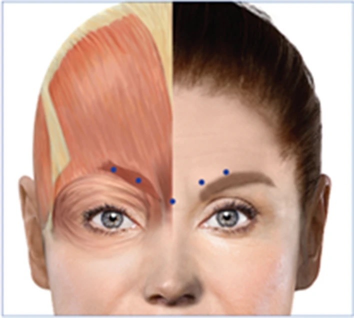
Figure : OnabotulinumtoxinA injection sites. This figure was modified from Blumenfeld and colleagues60 under a Creative Commons license (CC BY-NC 4.0).
Discussion
The facial feedback effect states that when we contract or flex relevant muscles to create an emotional expression (e.g., happy or angry), it can help identify and experience the reflected emotion, even in the absence of an emotional face as a stimulus. There is evidence that signaling between the emotional centers of the brain and facial muscles is bidirectional which contributes to a neural circuit involved in the processing of emotions. The activity of the corrugator muscle is detected through the facial nerves that innervate the proprioceptive fibers of the optic branch of the trigeminal nerve.
The trigeminal mesencephalic nucleus feeds the locus coeruleus and the amygdala which has direct connections with the prefrontal cortex, both structures critical for emotional regulation . The amygdala responds to emotional valence , often responding strongly to fear and arousal, but also when the specificity and differentiation of the emotion has self-relevance or a strong relationship to one’s goals. Therefore, deactivation of the glabellar region may have a downstream effect on the neuroanatomical circuitry involved in the processing of emotional faces.

Figure: Neuroanatomical circuit involved in the processing of emotional faces. The orange lines represent the sensory trigeminal nerve, which innervates the brain stem and synapses on the trigeminal nucleus. The blue lines highlight the flow of information along key regions of the circuit. Sensory neurons of the trigeminal nucleus caudalis have reciprocal connections with sensory and limbic structures and are often monosynaptic. These include trigemino-amygdala, trigemino-hypothalamus, trigemino-thalamus, and trigemino-locus coeruleus connections. Skull layers: white, scalp; rose, periosteum; gray, bone; blue, meninges (dura mater, arachnoid mater, pia mater); SpV: spinal tract of the trigeminal nucleus; SSN: superior salivary nucleus; VPM: ventral posteromedial nucleus. This figure was created in Adobe Illustrator.
Conclusions The present results provide additional evidence that neuromuscular feedback from the creation of an emotional expression can influence activity in two key regions for the processing of emotional faces: the amygdala and the fusiform gyrus. Inhibition of muscles in the glabellar region prevented frowning and reduced the creation of smiling or happy expressions, resulting in alterations in amygdala activity for both happy and angry faces. Increased amygdala activity may reflect compensatory processes during emotional processing that are activated when facial feedback is modulated. While much more remains to be explored regarding the role of facial feedback in the activity of the amygdala and fusiform gyrus, as well as other regions involved in the neuroanatomical circuitry for processing emotional faces, |
Clinical Trials.gov registration number: NCT03373162.


