Clinical vignette A 20-year-old woman with no medical history presents to the emergency department with “abnormal results” on a CT scan. She was feeling well until about 2 weeks ago when she started having fatigue, malaise, loss of appetite, and abdominal discomfort. She consulted her primary care physician, who ordered an outpatient CT scan of her abdomen and pelvis, and upon obtaining results showing splenomegaly , sent her to the emergency department for evaluation. |
Vital signs :
- FC 108 x’
- BP: 118/78 mm Hg
- FR: 20 x’
- Temperature: 38°
- SpO2 100% in room air
On physical examination , he has a scaphoid abdomen with a palpable mass on the left side extending 8 cm beyond the rib cage, consistent with splenomegaly. She appears fatigued and tachycardia, but the rest of her physical examination is normal.
What is the approach to the patient with splenomegaly?
Diagnosis : splenomegaly of unclear etiology.
Physiology:
The spleen is responsible for the removal of damaged red blood cells and plays an important role in the immune system by filtering pathogens from the blood and initiating immune reactions by bringing those foreign antigens into contact with specialized cells of the immune system.
The spleen plays an important role in hematopoiesis (production of blood cell lineages) during fetal development, but can become a site of extramedullary hematopoiesis in pathological states.
Pathophysiology:
A pathologically enlarged spleen, identified on examination or imaging, warrants evaluation to determine the underlying etiology.
The definition of splenomegaly varies, but most agree that a spleen larger than 10-13 cm in any direction is enlarged, although slightly larger spleens, up to 14 cm, may be physiological in tall people. A spleen measuring more than 20 cm is considered massive splenomegaly .
The three most common causes of splenomegaly are:
- Liver disease (33%)
- Hematologic malignancy (27%)
- Infection (23%)
Medical history and examination:
Important historical elements to obtain:
Age of the patient : For example, a child presenting with splenomegaly may be a new diagnosis of sickle cell disease, while an older person may be a new diagnosis of cancer.
Duration of symptoms
Presence of symptoms suggesting anemia or thrombocytopenia , including easy bruising/bleeding, difficulty breathing, fatigue.
Presence of symptoms that may suggest malignancy or infection, including weight loss, night sweats, lymphadenopathy, or fever
Past medical history of heart disease, liver disease, sickle cell disease, autoimmune disease, or cancer or recent trauma.
Physical examination technique to identify splenomegaly:
Have the patient lie down or in the right lateral decubitus position with arms at sides, then slowly palpate the left side starting at the rib cage and moving caudally to the pelvis to locate the lower border of the spleen, which can vary greatly with respiration. .
The use of bedside ultrasound in addition to physical examination significantly increases sensitivity in identifying splenomegaly.
The presence of splenomegaly may be missed on examination for multiple reasons:
- The spleen is enlarged but still too small to be palpated successfully; some articles suggest that the spleen must be enlarged by at least 40% to be palpable below the rib cage.
- The spleen is significantly enlarged and the lower edge of the spleen is in the pelvis.
- The patient’s body constitution may limit the examination.
Differential diagnosis:
The differential diagnosis of splenomegaly is broad and can be divided into 3 main categories:
1) Congestion due to increased vascular pressure.
2) Increase in size due to hemolysis of blood.
3) Infiltration by other cells/material.
Differential diagnosis for splenomegaly related to increased vascular pressure:
- Cirrhosis
- Congestive heart failure
- Thrombosis of the splenic vein, portal vein or splanchnic vein
- Vaso-occlusive crisis secondary to sickle cell disease leading to splenic sequestration
Differential diagnosis for splenomegaly related to blood hemolysis:
- Autoimmune hemolytic anemia
- Seen in patients with other autoimmune disorders, such as SLE, rheumatoid arthritis, Sjogren’s, Hashimoto’s thyroiditis, Felty syndrome
- immune thrombocytopenia
- Hemolytic uremic syndrome
- Thrombotic thrombocytopenic purpura
Differential diagnosis for splenomegaly related to cell/material infiltration
- Infiltration by cancer cells : primary malignancy (splenic marginal zone lymphoma), hematological cancers (ALL, CLL, CML, polycythemia vera) or metastasis (most commonly melanoma, breast and lung cancer).
- Infiltration by blood/lymphatic cells in response to infection: EBV, CMV, HIV, mycobacteria, brucellosis, babesiosis, bartonella, histoplasmosis, leptospirosis, malaria, visceral leishmaniasis.
- Amyloidosis
- Sarcoidosis
- Gaucher disease
- Other conditions
- splenic hemangioma
- splenic hemangiosarcoma
- Traumatic injury resulting in splenic hematoma
Emergency evaluation:
Given the wide differential for splenomegaly, evaluation in the emergency department typically includes obtaining the initial workup and determining whether the patient needs to be admitted for further workup or discharged with outpatient follow-up.
Obtain a lab profile that includes:
- Complete blood count with differential and smear.
- Liver function tests.
- Serological tests for possible infections.
- Hemolysis labs including lactate dehydrogenase (LDH), unconjugated bilirubin (indirect bilirubin), haptoglobin, and reticulocyte count.
Ultrasound
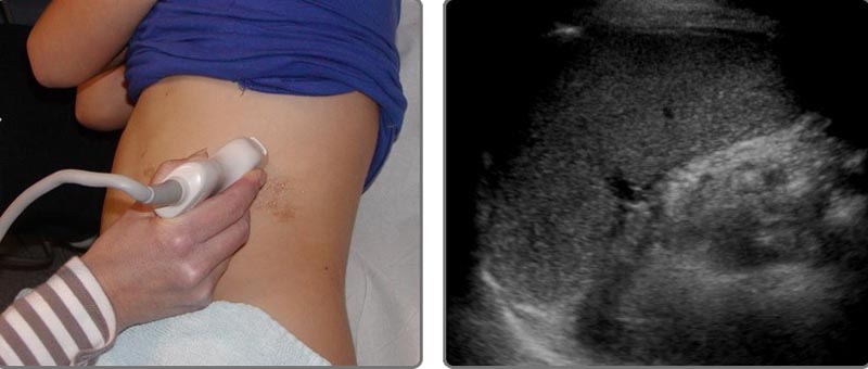
Intercostal examination plane. Note that the transducer has been rotated so that it is parallel to the rib space. Image of a normal spleen. Source: ULTRASOUNDPAEDIA https://ultrasoundpaedia.com/spleen-normal/
Clinical management:
A person with splenomegaly who appears ill should be admitted for expedited workup of the underlying etiology and consideration of splenic rupture.
A well-appearing person with atraumatic splenomegaly can be reasonably discharged with outpatient primary care and hematologic follow-up.
Management varies greatly depending on the etiology of splenomegaly. For example, admission and resuscitation for splenic sequestration due to sickle cell vaso-occlusive crisis versus discharge with supportive care and precautions in the infectious mononucleosis patient with mild splenomegaly.
Complications:
Splenic rupture is a feared and life-threatening complication of splenomegaly; Therefore, it is generally recommended to avoid contact sports to reduce this risk.
Cytopenia and immune dysfunction related to cell sequestration within the spleen can lead to bleeding and infection.
Main points to remember:
|


