Contrastive learning and subtyping of post-COVID-19 lung CT images Summary Patients who have recovered from the novel coronavirus disease 2019 (COVID-19) may experience a variety of long-term symptoms. Since the lung is the most common site of infection, pulmonary sequelae may occur persistently in COVID-19 survivors. To better understand the symptoms associated with lung function decline in post-COVID-19 patients, we aimed to build a deep learning model that performs two tasks: differentiating post-COVID-19 subjects from healthy subjects and identifying post-COVID-19 subjects. COVID-19 subtypes, based on latent representations from lung computed tomography (CT) scans . CT scans of 140 post-COVID-19 subjects and 105 healthy controls were analyzed. A new learning model was developed by introducing a lung volume transformation to learn latent features of disease phenotypes from inspiration and expiration CT scans of the same subjects. The model achieved 90% accuracy for differentiating post-COVID-19 subjects from healthy controls. Two groups (C1 and C2) with distinct characteristics were identified among post-COVID-19 subjects. C1 exhibited greater air trapping caused by small airway disease (4.10%, p = 0.008) and the diffusing capacity of %predicted carbon monoxide (%predicted DLCO, 101.95%, p < 0.001), while C2 presented a decrease in lung volume (4.40 l, p < 0.001) and an increase in ground glass opacity (GGO%, 15.85%, p < 0.001). The contrastive learning model is able to capture the latent characteristics of two post-COVID-19 subtypes characterized by air trapping due to small airway disease and airway-associated interstitial fibrotic patterns , respectively. The discovery of post-COVID-19 subtypes suggests the need for different management and treatments of the long-term sequelae of post-COVID-19 patients. |
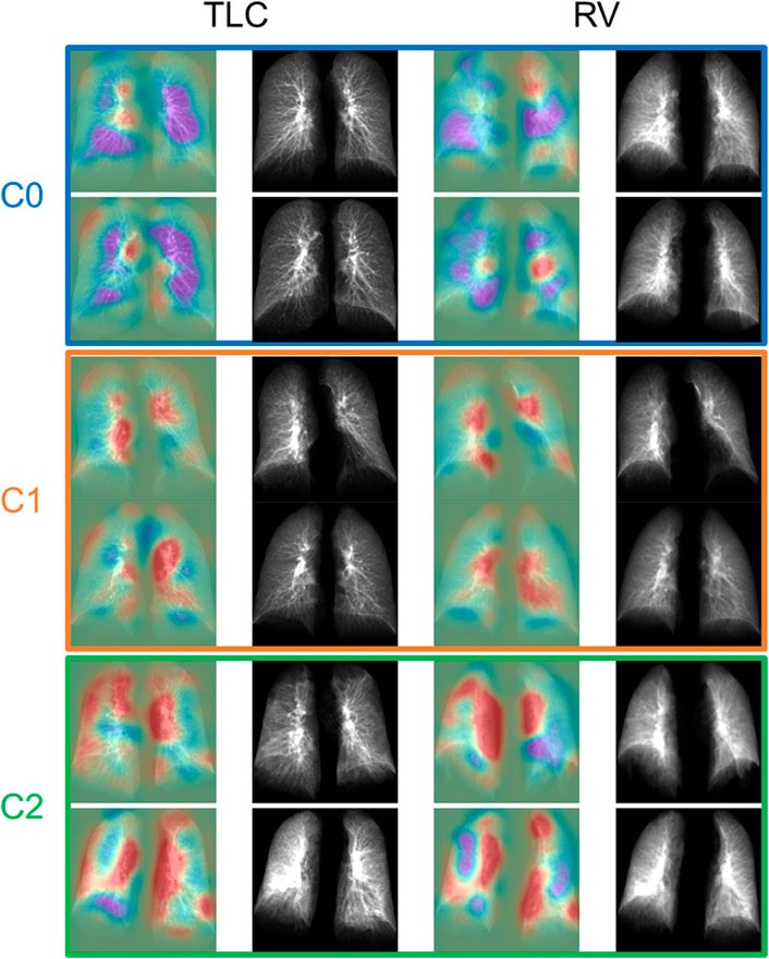
The TLC and VR images of the representative subjects of each group. The first and third columns showed the activation maps indicating the regions important for determining whether the subjects were post-COVID-19 subjects (red) or control subjects (purple).
Comments
For patients dealing with persistent respiratory symptoms of the new coronavirus, a chest X-ray can’t reveal much. Two-dimensional (2D) scans simply cannot distinguish compromised lung function. For this diagnosis, a more three-dimensional (3D) CT technique is needed.
However, many medical clinics in the United States do not have CT machines, leaving long COVID patients with little information about their lung function.
That may change. In a new study, researchers at the University of Iowa developed what is called a contrastive learning model . This model “learns” from composite 2D images constructed from 3D CT images to detect compromised lung function in long COVID patients. Another technique, called transfer learning , then transmits lung diagnostic information from a CT scan to a chest X-ray, allowing the chest X-ray machine to detect abnormalities as if those patients had used a CT scan.
In the study, the researchers showed how their contrastive learning model could be applied to detect small airway disease , which is an early stage of compromised lung function in long COVID patients. Of patients with long COVID, the models were advanced enough to distinguish the severity of compromised lung function, separating those with small airway disease from those with more advanced breathing problems.
“The new element of the model is to take information from 3D CT scans that show lung volume and transfer that information to a model that will show these same features in 2D images,” say Ching-Long Lin, Edward M. Mielnik and Samuel R Harding professor and chair of the Department of Mechanical Engineering at the Iowa College of Engineering. “Doctors could use chest x-rays to detect these findings. “That’s the broader perspective.”
The researchers based their model on CT scans of 100 people who were infected with the original strain of COVID and came to UI Hospitals & Clinics to be diagnosed with respiratory problems between June and December 2020. Many of these long COVID patients had illness of small airways, a diagnosis reported by Alejandro Comellas, clinical professor of internal-pulmonary medicine, intensive care and occupational medicine, in an article published last March in the journal Radiology.
Small airway disease affects a network of more than 10,000 tubes at the nexus in the lung where oxygenated air mixes with blood to be transported throughout the body. People with small airway disease have many of these blood vessels constricted, which limits the exchange of oxygen and blood in the lungs and prevents breathing in general.
Lin and his team collected data points at two intervals on CT scans of the lungs: when the patient inhaled and when he exhaled. The researchers compared their results to a control group that had not contracted the virus while creating the contrastive learning model.
“Our models successfully identified decreased lung function in patients with long COVID compared to those who had not contracted the virus,” says Lin, whose expertise is in machine learning and computational fluid and particle dynamics simulation.
Lin’s team advanced the model so that it could separate patients with small airway disease from those with more advanced complications, such as emphysema.
“The study independently demonstrated that post-COVID patients have two types of lung lesions (small airway disease and fibrosis/inflammation of the lung parenchyma) that are persistent after they have recovered from their initial SARS CoV infection.” 2,” says Comellas, co-author of this study.
“Chest X-rays are affordable, while CT scans are more expensive and not as accessible,” Lin adds. “Our model can be improved further, and I think there is potential for it to be used in all clinics without having to purchase expensive imaging equipment such as CT scanners.”
The authors note that the study is limited, in part because the sample size is small and the patients are from a single medical center. A larger sample size, they write, may uncover more variations in lung function stemming from long COVID.
The study, "Contrastive learning and subtyping of post-COVID-19 lung CT images," was published in the journal Frontiers in Physiology . Co-authors, all from Iowa, include Frank Li, Xuan Zhang, Eric Hoffman and Tianbao Yang.
The National Heart, Lung, and Blood Institute, a branch of the U.S. National Institutes of Health; and the U.S. Department of Education funded the research.


