It is usually diagnosed clinically and the goal of treatment is to clear the rash and prevent the spread of the infection to other people. Good skin hygiene measures and topical antiseptic treatment are usually appropriate.
Antibiotics should only be used in specific circumstances, and if necessary, oral administration is almost always preferable to topical, unless the infection is very localized.
Impetigo, also known as "school ulcers ," is a contagious bacterial skin infection.
Key points for practice:
|
Impetigo can affect people of any age, but it occurs most often in young children (i.e., ages two to six). Staphylococcus aureus and Streptococcus pyogenes , alone or together, are the most common causes of impetigo.
Impetigo can occur on an area of previously healthy skin or at the site of minor trauma that disrupts the skin barrier, such as a scrape, scrape, or eczema.
Impetigo is highly contagious and can be transmitted through direct contact, often spreading quickly through families, daycares, or schools.
Impetigo is most common in:
- Hot and humid climate.
- Poor hygiene conditions, e.g. overcrowding or close physical contact, e.g. e.g. Contact sports.
- People who have skin conditions or experience trauma that disrupt the normal skin barrier, e.g. eczema, scabies, fungal skin infections, abrasion, insect bites.
- People with diabetes mellitus.
- Immunocompromised people.
- People who use intravenous drugs.
There are two types of impetigo: bullous and non-bullous.
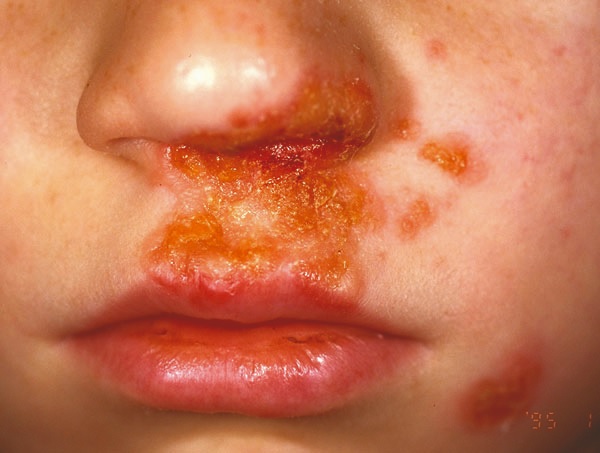
Nonbullous impetigo (Figure 1) is the most common variant, and is usually caused by S. aureus , but in some cases it can be caused by S. pyogenes . The lesions begin as a vesicle that ruptures and the contents dry out to form a gold. -colored plaque on the skin. These lesions are usually between 1 and 2 cm in diameter and most commonly affect the face (especially around the mouth and nose) and extremities. Fever and regional lymphadenopathy may appear.
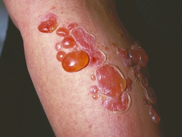
Bullous impetigo ( Figure 2) is caused by S. aureus alone and accounts for approximately 10% of cases, and is most commonly seen in infants. It is characterized by larger fluid-filled blisters that break less easily than non-blistering blisters. Impetigo, which leaves a yellowish-brown crust. Systemic signs of infection, such as fever and lymphadenopathy, are more likely to occur, and the trunk is more likely to be involved.

Figure 1. Non-bullous impetigo. Image provided by DermnetNZ

Figure 2 . Bullous impetigo. Image provided by DermnetNZ
Ecthyma is a form of deep tissue impetigo. It is characterized by scabbed ulcers beneath which "holey" appearance ulcers form. It is more common in children, the elderly and immunosuppressed or in conditions of poor hygiene and hot, humid climates. Treatment follows the same guidelines as impetigo, but oral antibiotics are usually required.
Impetigo is usually diagnosed clinically
Impetigo can be diagnosed by clinical examination and initial treatment decisions are rarely based on the results of skin swabs. Swabs may be necessary for people with recurrent infections, treatment failure with oral antibiotics or when there is an outbreak in the community and the cause needs to be identified. For people with recurrent impetigo, nasal swabs can identify nasal carriage of staph requiring specific treatment.
Impetigo treatment
Key points:
|
Topical antiseptic is the initial treatment for localized patches of impetigo.
A topical antiseptic, e.g. E.g. 1% hydrogen peroxide or 10% povidone-iodine, applied two to three times daily, is the first-line treatment for uncomplicated localized nonbullous impetigo (e.g., three or fewer lesions). / groups) should be removed with warm water before applying any topical preparation. Remind parents/caregivers to wash hands before and after application.
Five days of topical antiseptic treatment is usually sufficient to treat impetigo. This may be increased to seven days depending on the severity and number of injuries.
The use of topical antibiotics is discouraged
Topical antibiotics are rarely indicated for use in skin infections due to bacterial resistance and the possibility of contact dermatitis. However, there may be some cases where a topical antibiotic may be considered to treat a small, localized area of impetigo, as if it were topical impetigo. the antiseptic is not suitable (e.g., impetigo around the eyes) or has been ineffective.
Fusidic acid should be prescribed unless antibiotic sensitivity (if known) indicates resistance. Mupirocin is reserved for the treatment of MRSA.
Impetigo caused by methicillin-resistant S. aureus (MRSA)
The prevalence of impetigo caused by methicillin-resistant S. aureus (MRSA) is unknown , but is likely increasing. In August 201, 956 laboratory isolates of MRSA were reported in New Zealand, equivalent to a period prevalence rate of 19.9 MRSA patients per 100,000 population. This is double the 2009 isolates, but rates have remained relatively stable over the past four years.
Some community strains of MRSA show increasing resistance to fusidic acid, while resistance rates to mupirocin are decreasing concurrently with decreasing dispensing rates. Oral trimethoprim + sulfamethoxazole, tetracyclines or clindamycin are usually effective against MRSA.
Oral antibiotics should be used for multiple lesions or if topical treatment is ineffective. Oral antibiotics are recommended to treat patients with more than three to five lesions/clusters, bullous impetigo, systemic symptoms, or when topical treatment is ineffective. Flucloxacillin is the first-line choice as it is effective against S. aureus and group A streptococci.
Trimethoprim + sulfamethoxazole or erythromycin may be used if MRSA is present or if the patient is allergic or intolerant to flucloxacillin. Cephalexin is another option if flucloxacillin is not tolerated.
A five-day course of oral antibiotics is usually sufficient, but may be increased to seven days depending on the severity and number of lesions. If treatment is not successful after this time, medication adherence should be checked and swabs may be taken to check for sensitivities.
Prevention of recurrent impetigo infections
Recurrent infection may result from nasal carriage of causative microorganisms, close contact with others, or colonization by fomites, e.g. sheets, towels and clothes that can be shared.
If a nasal carrier is suspected , a nasal swab is required to confirm this and establish antibiotic susceptibility. A topical antibiotic should be applied inside each nostril, three times a day for seven days. The choice of antibiotic will be based on sensitivities (from the swab result).
All household members and close contacts should also receive treatment.
Complications of impetigo
- Poststreptococcal glomerulonephritis , which can lead to kidney failure, is a rare complication of streptococcal impetigo. Treatment of impetigo may not prevent susceptible people from developing this complication. The prevalence of poststreptococcal glomerulonephritis is highest in primary school-aged children, especially boys. and people of Māori and Pacific descent.
- Scarring may occur in people with more severe impetigo, when the lesions extend deeper into the dermis. In milder cases of impetigo, healed lesions can cause changes in skin pigmentation; however, this should resolve over time.
- A soft tissue infection such as cellulitis may occur .
- Staph scalded skin syndrome is characterized by red, blistered skin that leaves a burn-like area once the lesions have broken down. Children under five years of age, particularly newborns, immunocompromised people, or people with kidney failure, are at higher risk for this complication.
- Streptococcal toxic shock syndrome is a rare complication of impetigo. It is most commonly seen in healthy people ages 20 to 50, although children, immunocompromised people, and the elderly are more susceptible to impetigo. Symptoms include fever, rash, hypotension, and erythematous rash.
- Rheumatic fever is rarely related to skin infections; However, it can occur when group A strep found on the skin moves into the throat.


