
1. Sun exposure plays an important role in the pathogenesis of melanoma
More than 90% of melanomas in the 3 most common genetic subtypes (BRAF, RAS, NF1) have a substantial ultraviolet signature.1 Clinically, melanomas appear most frequently at sites of chronic (face, neck) or intermittent sun exposure (i.e., trunk, legs) such as superficial spread (Figure 1), nodular melanoma, or lentigo maligna.
Figure 1:
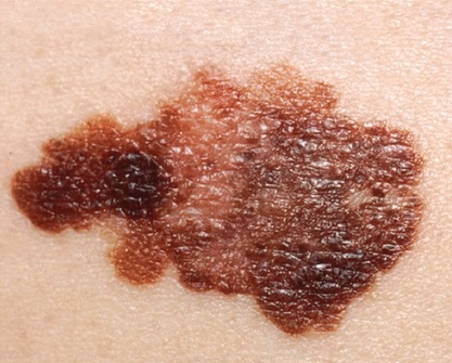
ABCDE criteria (asymmetric shape, irregular border, color variation, diameter > 6 mm [approximately the size of a pencil eraser], and evolution [noted in history and in comparison with previous size]) of a typical superficial melanoma that occurs characterized as a plate with flat and raised areas. Image obtained from the National Cancer Institute.
2. Melanomas also occur in sites of minimal sun exposure
Genome sequencing of acral (on the palms and soles of the feet) and mucosal melanomas has shown that, although sun exposure may play a role, it is not the main mutational driver.2 These melanomas may share a pathogenesis similar to that of other non-cutaneous malignancies.
3. The mitogen-activated protein kinase pathway is involved in almost all melanomas
This pathway stimulates cell growth and survival. BRAF, RAS, and NF1 mutations are part of this pathway and account for 50%, 25%, and 15% of all melanomas, respectively.1 Genome-wide studies have helped identify important immunohistochemical markers for diagnosis (e.g. e.g., unprogrammed cell death 1 [anti-PD-1], T cell-associated protein 4 [anti-CTLA4] 3, as well as adjuvant immunotherapies and targeted therapies (i.e., BRAF or MEK inhibitors) to improve the survival of patients with advanced melanoma.4
4. Of all melanomas, 10% are amelanotic or hypopigmented and can be difficult to diagnose 5
They are more common in patients with Fitzpatrick type I skin and chronic sun damage (actinic keratoses), and are located in sites exposed to the sun (for example, face, neck, arms, hands).5 The differential diagnosis of the evolution of red or pink. Macules, plaques, or nodules should include amelanotic melanoma, especially in the patients and locations mentioned above. KIT, a tyrosine kinase inhibitor, is frequently mutated in amelanotic melanoma.
5. Any lesion suspicious for melanoma should be referred to dermatology.
A pigmented lesion with any of the ABCDE criteria (Figure 1) should raise suspicion of melanoma. Definitive treatment is wide local excision with appropriate margins. In cosmetically sensitive areas (for example, the face), Moh surgery, in which thin layers of the tumor are sequentially removed until only cancer-free tissue remains, may be preferable.
Bibliographic references
- Cancer Genome Atlas Network. Genomic classification of cutaneous melanoma. Cell 2015;161:1681–96.
- Hayward NK, Wilmott JS, Waddell N, et al. Whole-genome landscapes of major melanoma subtypes. Nature 2017; 545:175–80.
- Massi D, Simi L, Sensi E, et al. Immunohistochemistry is highly sensitive and specific for the detection of NRASQ61R mutation in melanoma. Mod Pathol 2015;28:487–97.
- Long GV, Hauschild A, Santinami M, et al. Adjuvant dabrafenib plus trametinib in stage III BRAF-mutated melanoma. N Engl J Med 2017;377:1813–23.
- Wee E, Wolfe R, Mclean C, et al. Clinically amelanotic or hypomelanotic melanoma: anatomic distribution, risk factors, and survival. J Am Acad Dermatol 2018;79:645–51.e4.


