Clinical vignette A 73-year-old man presented with a mildly painful, nonpruritic lesion on the left ear (Figure 1) that had been present for 6 months, and he noted discomfort when sleeping on a firmer pillow on the left side. There was no history of trauma or excessive sun exposure. The patient periodically removed the scab, which always reappeared. He reported sleeping exclusively on his left side. The examination revealed a 3 mm lesion in the upper pole of the helix of the ear without erythema. There was a central scab over an ulcerated nodule without discharge or bleeding. The nodule was painful on palpation. |
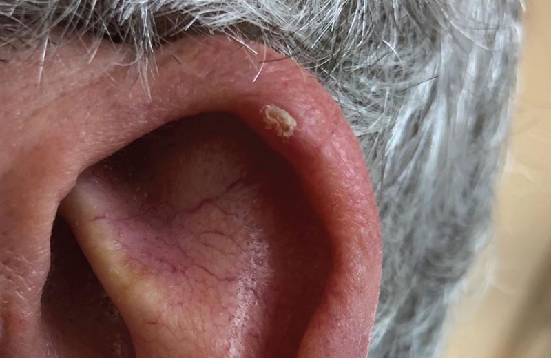
Figure 1 : On presentation, the lesion on the helix of the patient’s left ear was mildly painful and nonpruritic, with a central scab over an ulcerated nodule.
Approach
Based on the appearance of the lesion and the patient’s history of discomfort after switching to a firmer pillow, the lesion was diagnosed as nodular chondrodermatitis of the helix (CNH).
Our patient was advised to avoid pressing on the ear and sleep on the opposite side.
Chondrodermatitis nodularis helicis
Classically, Chondrodermatitis nodularis helicis (CNH) is a benign, painful inflammatory nodule or papule on the helix or antihelix of the ear that is sensitive to touch or pressure. Typical lesions are unilateral, 4 to 6 mm in size, and consist of an ulcerated nodule with a central crater. Crusting may or may not be present, and some lesions may have a cystic appearance.
Chondrodermatitis nodularis helicis (CNH) is most common in light-skinned middle-aged to older men and has a variable male-to-female ratio. The etiology of CNH is multifactorial and may result from thinning of the skin and cartilage seen with aging and pressure degeneration of cartilage. CNH can often cause sleep disorders when patients continue to sleep on the affected side. Affectation of the right side may be more common.
Differential diagnosis may include actinic or seborrheic keratosis, basal cell or squamous cell carcinoma, gouty tophi, and keratoacanthoma.
Patients are often referred to specialists for biopsy evaluation. However, taking a detailed history, specifically about sleep patterns, in combination with the location of the injury should help establish the proper diagnosis.
Biopsy should be performed when the diagnosis is uncertain, when there is a history of skin cancer or sun-damaged skin, or when the lesion does not respond to non-invasive interventions.
Treatment
As the pathophysiology of Chondrodermatitis nodularis helicis (CNH) is believed to be similar to that of pressure ulcers , treatment usually consists of conservative measures, such as avoiding or relieving pressure by sleeping on the contralateral side, plugging the ear with sponges or foam and use of a donut-shaped pillow. Clinical response to these interventions may obviate the need for a biopsy.
Other non-invasive therapies include intralesional steroid injections, topical nitroglycerin gel, cryotherapy, carbon dioxide laser therapy, or photodynamic therapy that uses a light source to improve blood flow. Wedge resection should be considered when the lesion recurs despite multiple attempts at less invasive interventions.
Resolution of the clinical case
At follow-up 2 months later, our patient reported significant improvement in symptoms, further solidifying the diagnosis of Chondrodermatitis nodularis helicis (CNH). The lesion was significantly smaller and the patient was informed that CNH can frequently recur and that other treatments may be needed.


