
Highlights
|
CHICAGO
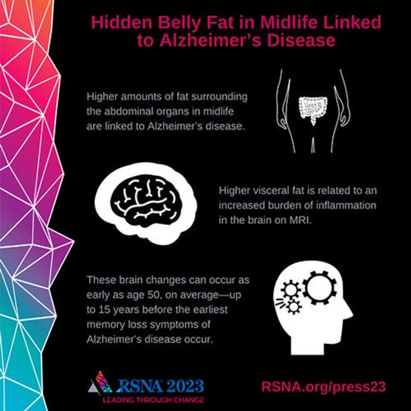
Greater amounts of visceral abdominal fat in middle age are linked to the development of Alzheimer’s disease, according to research to be presented next week at the annual meeting of the Radiological Society of North America (RSNA). Visceral fat is the fat that surrounds the internal organs deep in the abdomen. Researchers found that this hidden abdominal fat is linked to changes in the brain up to 15 years before the first memory loss symptoms of Alzheimer’s disease occur.
According to the Alzheimer’s Association, there are more than 6 million Americans living with Alzheimer’s disease. By 2050, this number is expected to increase to almost 13 million. One in five women and one in 10 men will develop Alzheimer’s disease during their lifetime.
To try to identify Alzheimer’s risks earlier, researchers evaluated the association between brain MRI volumes, as well as amyloid and tau uptake on positron emission tomography (PET) scans, with body mass index (BMI). , obesity, insulin resistance and abdominal adipose tissue in a cognitively normal middle-aged population. Amyloid and tau are thought to interfere with communication between brain cells.
"Although there have been other studies linking BMI to brain atrophy or even an increased risk of dementia, no previous study has linked a specific type of fat to the actual Alzheimer’s disease protein in cognitively normal people," the study said. Study author Mahsa Dolatshahi, MD. MPH, postdoctoral researcher at the Mallinckrodt Institute of Radiology (MIR) at Washington University School of Medicine in St. Louis. "Similar studies have not investigated the differential role of visceral and subcutaneous fat, especially in terms of Alzheimer’s amyloid pathology, already in middle age."
For this cross-sectional study, researchers analyzed data from 54 cognitively healthy participants, ages 40 to 60, with an average BMI of 32. Participants underwent glucose and insulin measurements, as well as insulin tolerance tests. glucose. The volume of subcutaneous fat (fat under the skin) and visceral fat was measured by abdominal MRI. Brain MRI measured the cortical thickness of brain regions affected by Alzheimer’s disease. PET was used to examine disease pathology in a subset of 32 participants, focusing on the amyloid plaques and tau tangles that accumulate in Alzheimer’s disease.
The researchers found that a higher proportion of visceral and subcutaneous fat was associated with greater uptake of the PET tracer amyloid in the precuneus cortex , the region known to be affected early by amyloid pathology in Alzheimer’s disease. This relationship was worse in men than in women. The researchers also found that higher measurements of visceral fat are linked to a greater load of inflammation in the brain.
"Several pathways are suggested to play a role," Dr. Dolatshahi said. " Inflammatory secretions from visceral fat, unlike the potentially protective effects of subcutaneous fat, can lead to inflammation in the brain, one of the main mechanisms contributing to Alzheimer’s disease."
Senior author Cyrus A. Raji, M.D., Ph.D., associate professor of radiology and neurology and director of neuromagnetic resonance imaging at MIR, said the findings have several key implications for earlier diagnosis and intervention.
"This study highlights a key mechanism by which hidden fat may increase the risk of Alzheimer’s disease," he said. "This shows that such brain changes occur at an early age of 50 years, on average, up to 15 years before the first symptoms of Alzheimer’s memory loss occur."
Dr. Raji added that the results may point to visceral fat as a treatment target to modify the risk of future brain inflammation and dementia.
"By going beyond body mass index and better characterizing the anatomical distribution of body fat on MRI, we now have an exceptionally better understanding of why this factor may increase the risk of Alzheimer’s disease," he said.
Additional co-authors are Paul K. Commean, BEE, Joseph E. Ippolito, MD, Ph.D., Tammie LS Benzinger, MD, Ph.D. and John C. Morris, MD.


