Key points • Cellulitis is a common skin infection, typically presenting with poorly circumscribed unilateral erythema, warmth, and tenderness, which has many clinical mimics. • A thorough history and clinical examination can narrow the differential diagnosis of cellulitis and minimize unnecessary hospitalization. • Treatment should be dictated by the patient’s history and risk factors, clinical presentation, and the most likely culprit pathogen, optimizing antibiotic administration. |
Cellulitis is a common infection of the skin, dermis, and subcutaneous tissue.
From 1998 to 2013, hospitalizations for cellulitis doubled and costs increased almost 120%. Cellulite can be difficult to identify given its numerous clinical mimics and the lack of a gold standard diagnostic test. The impossibility of confirming potential microbiological causes can further complicate management.
| Pathophysiology |
Cellulitis is a skin infection usually precipitated by the entry of bacteria through a crack in the skin barrier. The most common cause of nonpurulent cellulitis is Streptococcus pyogenes . This cellulitis is characterized by not presenting pustules or purulent drainage.
Nonpurulent cellulitis does not have a cultivable wound. Staphylococcus aureus can also cause nonpurulent cellulitis and is the most common cause of purulent cellulitis . Cellulitis may be caused by methicillin-resistant (MRSA) or methicillin-susceptible (MSSA) S. aureus , which can be difficult to distinguish clinically without wound culture and blood tests. sensitivity, and has implications for antibiotic selection.
The incidence of MRSA has been increasing in communities, and many patients with MRSA infection do not have any risk factors. On the other hand, risk factors for MRSA colonization are: previous use of antibiotics, recent hospitalization or surgery, prolonged residence in a service, infection with the human immunodeficiency virus (HIV), use of injection drugs, incarceration, military service, sharing sports equipment and razors.
Other potential pathogens are rarer and should be considered based on the clinical context. Cellulitis at the bite site of a dog or cat may be due to Pasteurella , Neisseria or Fusobacterium , while the organisms to consider in human bites are Eikenella corrodens or Veillonello . Cellulitis in the setting of aquatic injury may include Vibrio , Aeromonas , or Mycobacterium .
In immunocompromised patients it is important to investigate the etiology when possible, including non-bacterial causes. Helicobacter cinaedi can cause cellulitis in patients with HIV infection or a recent history of chemotherapy. Patients with systemic lupus erythematosus are susceptible to Streptococcus pneumoniae cellulitis . The clinical history is important to elucidate what the causative organism of infectious cellulitis may be and for the selection of antibiotic treatment.
Predisposing factors for infectious cellulitis include advanced age, obesity, leg edema, and previous infectious cellulitis. Lymphedema in particular can harbor bacterial growth. A retrospective study of more than 165,000 hospital admissions for lymphedema or cellulitis found that 92% of cases were related to cellulitis.
Lymphedema, venous insufficiency, and vascular disease have been shown to be predictive of cellulitis recurrence.
Furthermore, disruption of the skin’s barrier function due to chronic wounds, infection, or trauma is an important modifiable risk factor whose appropriate management improves outcomes.
| Clinical presentation |
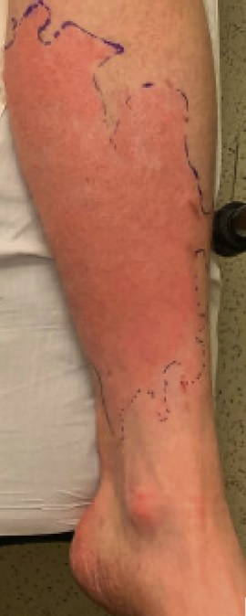
Cellulitis usually presents with poorly defined erythema, edema, pain on palpation, and heat of the affected skin.

Figure 1. Cellulitis of the lower extremities. Shiny, tender, warm erythematous plaque on the left lower extremity suggestive of cellulitis.
Erysipelas can be considered a type of cellulitis that affects the superficial dermis and presents with well-defined erythema. The clinical presentation of cellulitis is often distinguished by the presence or absence of purulence. It can be complicated by the formation of skin abscesses, a collection of pus enclosed in the subcutaneous space that may require surgical intervention.
Other findings may be lymphangitis, lymphadenopathy, vesiculation or blisters. Fever is sometimes present, with a very wide estimate of the incidence ranging from approximately 22% to 77%, depending on the clinical setting of the study performed.
Cellulitis can affect any area of the body, although the lower extremities are the most common site of infection in adults.
A retrospective study specifically evaluating lower extremity cellulitis developed the ALT-70 cellulite score and demonstrated that the clinical factors of unilateral cellulitis (+3, if true), age ≥70 years (+2, if true ), leukocytosis ≥10,000/µl (+1, if true), and tachycardia ≥9 0 beats/min (+1, if true) were predictive of true cellulitis.
The ALT-70 cellulite score has a >82% positive predictive value for predicting true cellulitis, if the calculated scores are ≥ 5. This may be a valuable tool for clinicians when evaluating lower extremity cellulite.
Cellulitis often does not present with outstanding features on laboratory evaluation, and blood tests are generally not required when evaluating patients with uncomplicated cellulitis or without comorbidities. Findings tend to be nonspecific and may show leukocytosis in less than 50% of patients while inflammatory markers are elevated. If the cellulitis is purulent, the 2014 Infectious Diseases Society of America (IDSA) guidelines recommend a Gram stain and culture.
Wound culture is not recommended in patients with nonpurulent cellulitis given the lack of a cultureable source from bare skin samples. Furthermore, the IDSA only recommends blood cultures in immunocompromised patients, malignancy, signs of systemic infection, or animal bites. In febrile patients with neutropenia, the acquisition of radiological images is recommended.
A retrospective study of patients with uncomplicated cellulitis found that most of them were evaluated by radiographs and blood cultures without meeting IDSA criteria. The evaluation had low clinical utility, rarely led to a change in treatment, and generated considerable unnecessary annual cost.
| Differential diagnosis |
Cellulite has many clinical mimics . The misdiagnosis rate of cellulite has been estimated at almost 30%, with some estimates as high as 74%. A thorough history and clinical examination can help distinguish cellulite from its clinical mimics. If patients with cellulitis do not improve with appropriate therapy or exhibit features other than cellulitis, such as bilateral or symmetrical findings, it is important to consider alternative diagnoses.
Stasis dermatitis is an inflammatory skin condition of the lower extremities that occurs in patients with chronic venous insufficiency. It is a common clinical mimic of cellulitis that can often be ruled out given its bilateral presentation in the absence of trauma, although it may rarely present unilaterally based on anatomical variation of the vein or leg injury. Improvement with compression and elevation of the legs and treatment with topical corticosteroids favor the diagnosis of stasis dermatitis, ruling out cellulitis.
Contact dermatitis is an inflammatory response of the skin to an irritant or allergen; Up to 80% of cases tend to be due to an irritant. A characteristic that distinguishes cellulitis from contact dermatitis is the symptom of itching, although in severe cases there may be pain. Although cellulitis and contact dermatitis may present with erythema, contact dermatitis may distribute in a geometric shape or pattern due to the triggering agent.
The etiology can be clarified by taking a thorough history investigating possible triggers, such as detergents, soaps, plants, or fragrances. If the etiology is allergenic, patch testing may clarify the offending agent. The pillars of management are elimination of the triggering agent, treatment of the skin with topical corticosteroids, and treatment of pruritus with antihistamines.
It is important to rule out necrotizing soft tissue infection (such as necrotizing cellulitis and necrotizing fasciitis), given its severity and high mortality rate. Patients with signs of systemic toxicity, rapidly progressive erythema or purpura, and disproportionate pain on physical examination require immediate surgical evaluation and antibiotic treatment.
Specifically, to detect necrotizing fasciitis when the initial clinical suspicion is not strong enough to warrant immediate surgical exploration , the Laboratory Risk Indicator for Necrotizing Fasciitis (LRINEC) score necrotizing fasciitis) incorporates C-reactive protein levels; leukocyte count, hemoglobin, sodium, creatinine and glucose. A meta-analysis demonstrated that a LRINEC ≥ 8 had a specificity of 94.9% for detecting necrotizing fasciitis.
| Differential diagnosis of cellulite |
| > Infection • Necrotizing soft tissue infection (necrotizing fasciitis). • Erythema migrans • Herpes zoster |
| > Non-infectious, inflammatory • Contact dermatitis • Sweet syndrome • Gout • Erythema nodosum |
| > Vascular • Stasis dermatitis • Deep vein thrombosis • Erythromelalgia |
| > Neoplastic • Carcinoma erysipelatoid |
| Treatment |
For several decades, systemic inflammatory response syndrome (SIRS) was commonly used to define the severity of sepsis and categorize the severity of cellulitis. The score incorporates temperature >38ºC or <36ºC; heart rate >90 beats/min; respiratory rate >20 breaths/min or partial pressure of carbon dioxide <32 mm Hg, and leukocytes >12,000/mm3, <4000/mm3, or >10% immature bands.
Mild cellulitis is characterized by no signs of systemic infection. Moderate cellulite is characterized by meeting 1 or 2 SIRS criteria. Cellulitis is severe if it meets ≥2 SIRS criteria plus hypotension, immunocompromise, or rapid progression.
Mild cellulitis is usually treated with oral antibiotics and severe cellulitis with intravenous antibiotics. Moderate cellulitis can be treated with oral or intravenous antibiotics depending on whether you meet 1 or 2 SIRS criteria. However, the Quick Sequential [sepsis-related] Organ Failure Assessment (qSOFA) score has become the new standard for sepsis risk assessment.
It is scored by respiratory rate ≥22 breaths/min; alteration of mental activity, which can be assessed with the Glasgow Coma Scale <15; and systolic blood pressure ≤100 mmHg. Given the limited literature evaluating qSOFA scores with cellulite outcomes, general principles for characterizing cellulite are based on sustained vital sign abnormalities.
Mild cellulitis can be characterized by not meeting qSOFA criteria while severe cellulitis is represented by a qSOFA score ≥2, which is associated with poor sepsis outcomes.
In addition to vital sign abnormalities, the therapeutic approach to cellulitis depends on its clinical presentation: nonpurulent, purulent without skin abscess, or purulent complicated with skin abscess. Cellulitis without skin abscess is generally managed with antibiotics while for skin abscess the management is surgical.
Antimicrobial stewardship involves using the narrowest spectrum of antibiotic activity necessary to treat cellulite.
The IDSA recommends starting treatment of uncomplicated cellulitis with oral antibiotics taking into account the most likely etiological agent. Unnecessary antibiotic coverage can promote organism resistance to the drugs, side effects, and increased costs. The empirical treatment of non-purulent cellulitis explains the presence of the most common pathogens, Streptococcus pyogenes and MSSA.
Patients with mild nonpurulent cellulitis who can tolerate oral therapy and have no risk factors for MRSA may be treated with oral antibiotics such as cephalexin, amoxicillin-clavulanic acid, or dicloxacillin.
Patients with a true allergy to penicillin can be treated with clindamycin. A randomized study of 153 patients with cellulitis without abscess demonstrated comparable cure rates in patients receiving cephalexin for empirical treatment of Streptococcus pyogenes and coverage of MSSA versus those treated with cephalexin and trimethoprim-sulfamethoxazole for additional empirical coverage of MRSA. Similarly, a larger randomized study of 496 patients with nonpurulent cellulitis demonstrated that the use of cephalexin and trimethoprim-sulfamethoxazole (TMP-SMX) compared with cephalexin alone did not result in higher rates of clinical resolution of cellulitis. .
Empiric antibiotic treatment against community acquired MRSA in patients with uncomplicated cellulitis does not appear to improve outcomes. Moderate nonpurulent cellulitis without signs of hypotension, immune compromise, or rapid deterioration can be treated with intravenous cefazolin or ceftriaxone, while severe nonpurulent cellulitis with any of these features requires broad-spectrum antibiotic coverage, such as vancomycin and piperacillin-azobactam. and additionally consider surgical evaluation for necrotizing fasciitis. Since purulent cellulitis without abscess is usually caused by S. aureus , antibiotics are recommended.
Selection depends on suspicion of MRSA. Although a wound culture and an antibiogram can determine the microbial cause, doctors are often faced with the need to perform empirical treatment, because cultures can take up to 5 days to provide a result.
Mild purulent cellulitis without MRSA risk factors can be treated similarly to nonpurulent cellulitis, with oral antibiotics such as cephalexin or dicloxacillin. Patients with suspected MRSA infection may be treated with oral antibiotics, to cover MRSA, such as trimethoprim-sulfamethoxazole or doxycycline. Although oral clindamycin has comparable efficacy to TMP-SMX in treating cellulitis, it is generally not recommended as a first-line agent in patients without penicillin allergy, given the risk of Clostridioides difficile infection.
The general principles also apply for moderate infection and severe purulent cellulitis. Moderate purulent cellulitis with low suspicion of MRSA can be treated with intravenous oxacillin or cefazolin. If suspicion of MRSA infection is high, intravenous vancomycin or clindamycin is preferable. Severe purulent cellulitis warrants extensive coverage of intravenous antibiotics and evaluation for necrotizing fasciitis. When clinical improvement appears, antibiotic coverage can be narrowed based on the antibiogram.
Purulent cellulitis with a drainable abscess is treated by incision and drainage. The ISDA does not recommend antibiotics for mild skin abscesses characterized by the presence of a single abscess that can be drained, in patients without signs of systemic infection or immunocompromise. However, 2 recent studies have suggested that, in addition to incision and drainage, higher cure rates could be achieved with adjuvant antibiotic therapy for MRSA.
A multicenter randomized controlled trial of 1247 patients seen in the emergency department and presenting with an abscess ≥2 cm in diameter determined that adjuvant treatment with TMP-SMX resulted in increased cure rates of approximately 7%, as well as higher cure rates. lower rates of subsequent surgical procedures by 5.2%, and similar rates of adverse effects.
Another multicenter randomized controlled study of 786 patients in the emergency department or outpatient setting presenting with an abscess ≤5 cm also demonstrated that adjuvant treatment with TMP-SMX or clindamycin resulted in higher cure rates of 12.8%. and 14.2%, respectively. In this trial, adjuvant clindamycin also had lower rates of recurrent infection within 30 days compared to placebo, at 5.4%. However, patients who received clindamycin had a nearly 10% higher rate of adverse events, such as non- C. difficile- associated diarrhea .
A meta-analysis investigating these studies and 2 smaller randomized controlled trials using adjuvant TMP-SMX replicated key findings. The adjuvant MRSA antibiotic group had a 7.4% higher cure rate and a 4.4% higher rate of antibiotic side effects, with no long-term sequelae. These results imply a modest benefit of treatment of uncomplicated abscesses treated with incision and drainage and adjuvant MRSA antibiotics. Clinical response dictates the duration of antibiotic therapy.
Evidence supporting the use of antibiotics for more than 5 days for uncomplicated cellulitis is lacking. For uncomplicated cellulitis, the authors recommend prescribing an initial course of antibiotics for 5 days with close follow-up within 2-3 days to ensure adequate clinical improvement. Lack of improvement may require a change in antibiotic coverage or reassessment of pseudocellulitis. In immunocompromised patients, the duration of treatment is 7-10 days although it is recommended up to 14 days.
The diagnosis of cellulite can be difficult and dermatological evaluation has become a useful consultation.
A randomized controlled trial demonstrated that dermatologists identified pseudocellulitis at a rate of 30.7% in patients with suspected cellulitis compared to a 5.7% rate by the primary team. Early dermatological consultation improved outcomes in patients with suspected cellulitis by identifying and managing clinical mimics, treating modifiable risk factors predisposing to cellulitis, and decreasing the duration of unwarranted antibiotic treatment.
Another study from the United Kingdom demonstrated that early dermatological consultation can be a cost-effective intervention by reducing inappropriate antibiotic use and hospitalization. Similar findings have been replicated in the US where, in patients with pseudocellulitis, dermatological consultation reduced rates of unnecessary antibiotic use by 74.4% and hospitalization by 85.0%, limiting exposure to antibiotics. antibiotics in more than 90,000 patients saving more than $210 million a year.
| Antibiotic cellulite treatment for coverage | |||
| Via | Antibiotic | Recommended dose | Comments |
| Streptococcus and coverage for MSSA | |||
| Oral | Amoxicillin–clavulanic acid | 875 mg 2 x day | __ |
| Cephalexin | 500 mg 4 x day | Adding TMP-SMX to Cephalexin for Empirical MRSA Coverage Does Not Offer Beneficial Clinical Outcomes | |
| Dicloxacillin | 250-500 mg 4 x day | Preferably oral to cover MSSA | |
| Penicillin VK | 250-500 mg 4 x day | __ | |
| Intravenous | Cefazolin | 1 g 3 x day | Alternative for patients with penicillin allergy without a history of immediate hypersensitivity reaction |
| Ceftaroline | 600 mg 2 x day | __ | |
| Ceftriaxone | 1-2 gx day | __ | |
| Imipenem | 500 mg 4 x day | Administered with cilastatin to prevent rapid inactivation | |
| Meropenem | 1 g 3 x day | __ | |
| Nafacillin or oxacillin | 1-2 gc/4h | Preferably parenteral agent to cover MSSA | |
| Penicillin G | 2-4 million units every 4-6 hours | __ | |
| Piperacillin-tazobactam | 3375 g 4 x day | Recommended use with vancomycin for empirical coverage of serious infections | |
| MRSA Coverage | |||
| Oral | Clindamycin | 300-400 mgf 4 x day | Risk of C. difficile infection Alternative for patients with penicillin allergy |
| Doxycycline or minocycline | 100 mg 2 x day | Variable antistreptococcal activity. It can be administered with amoxicillin to improve strep coverage. | |
| Linezolid | 600 mg 2 x day | Alternative for patients with allergy to ß-lactams High cost | |
| TMP.-SMX | 1-2 buy Forte 2 x day | __ | |
| Intravenous | Clindamycin | 600 mg 3 x day | __ |
| Daptomycin | 4 mg/kg x day | Alternative for patients who do not tolerate vancomycin | |
| Linazoid | 600 mg 2 x day | __ | |
| Telavancin | 100 mg/ x day | __ | |
| Tigecycline | 100 mg, then 50 mg 2 x day | __ | |
| Vancomycin | 150 mg/ 2 x day | Used with piperacillin-tazobactam or imipenem and meropenem for empirical coverage of serious infections | |
Summary Cellulitis is a common skin infection that has resulted in increased hospitalizations and costs. Although cellulite can be challenging to distinguish from its imitators, a thorough clinical examination can narrow the differential diagnosis and appropriately guide its management. Antibiotic selection is determined based on clinical presentation, patient risk factors, and the most likely causative microbial agent. Dermatologic evaluation has been associated with improved clinical outcomes and may help control this pervasive infection. Additional research is needed to improve the diagnosis and treatment of cellulite. |


